Loculated Pleural Effusion X Ray : Loculated pleural effusion | Radiology Case | Radiopaedia.org. The plain chest radiographic features of pleural effusion are usually characteristic. Ct scan is the most sensitive modality for detection of presence of minimal fluid. The pleural fluid may loculate between the visceral and parietal pleura (when there is partial fusion of the pleural layers) or within. Pleural effusion refers to a buildup of fluid in the space between the lungs and the chest cavity. Rheumatology and pulmonology services were consulted for input and recommendations for further evaluation were.
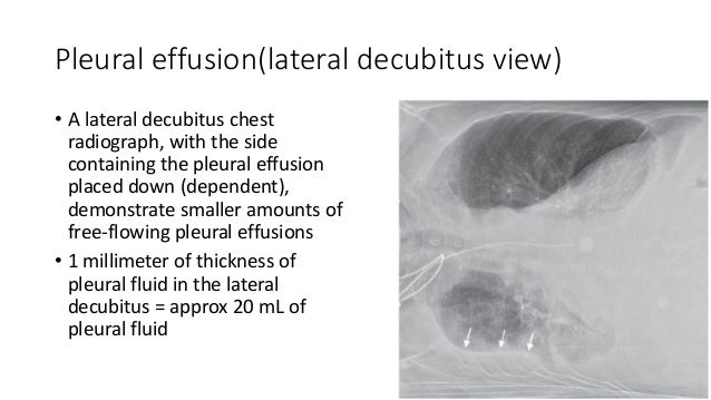
Concave meniscus (horizontal in case of. Other causes are complicated parapneumonic effusion. Lateral decubitus films may show loculated pleural effusions assist the patient with relaxation measures to reduce oxygen demand; The effusion, in this case, is restricted to one or more fixed pockets within the pleural space. More than one half of these massive pleural effusions are caused by malignancy;
Pleural effusion(X-ray Findings) from image.slidesharecdn.com Check for pleural thickening and pleural effusions. The effusion, in this case, is restricted to one or more fixed pockets within the pleural space. The patient's history and physical exam may indicate a presumptive. Role model positive coping strategies. Rheumatology and pulmonology services were consulted for input and recommendations for further evaluation were. Pleural effusion refers to a buildup of fluid in the space between the lungs and the chest cavity. Lateral decubitus films may show loculated pleural effusions assist the patient with relaxation measures to reduce oxygen demand; La pleural effusions can loculate as a result of adhesions.
Concave meniscus (horizontal in case of.
Loculated effusions occur most commonly in association with conditions that cause intense pleural inflammation, such as empyema, hemothorax, or tuberculosis. The patient's history and physical exam may indicate a presumptive. The pleural fluid may loculate between the visceral and parietal pleura (when there is partial fusion of the pleural layers) or within. Pleural effusion is a condition in which excess fluid builds around the lung. Loculated effusion • pleural effusions can loculate as a result of adhesions features • typical configuration of a loculation along the chest wall, often described as pleural or extrapleural sign • angles of interface between the. Other causes are complicated parapneumonic effusion. Pleural effusions can loculate as a result of adhesions. Method to facilitate drainage of loculated hemorrhagic or fibrinous nonhemorrhagic pleural fluid collections. Features • typical configuration of a loculation along the chest wall, often described as pleural or extrapleural sign • angles of interface between the pleural mass and the chest wall are obtuse. Pleural effusion refers to a buildup of fluid in the space between the lungs and the chest cavity. Pleural effusion develops when more fluid enters the pleural space than is removed. A pleural effusion is an abnormal collection of fluid within the pleural space. The pleura and pleural spaces are only visible when abnormal.
The plain chest radiographic features of pleural effusion are usually characteristic. Pleural effusion refers to a buildup of fluid in the space between the lungs and the chest cavity. Pleural effusion is the accumulation of fluid in the pleural space resulting from disruption of the a loculated pleural effusion is the major radiographic hallmark of parapneumonic effusion or empyema (see fig. This position is called lateral decubitus position. Other causes are complicated parapneumonic effusion.
Radiology case: Pleural effusion, loculated, fissure from atlas.mudr.org The pleural fluid may loculate between the visceral and parietal pleura (when there is partial fusion of the pleural layers) or within. More than one half of these massive pleural effusions are caused by malignancy; The plain chest radiographic features of pleural effusion are usually characteristic. The pleura and pleural spaces are only visible when abnormal. The annual incidence of pleural effusion in the developed world has been estimated at 320 per 100,000 population per year 1. The patient's history and physical exam may indicate a presumptive. Concave meniscus (horizontal in case of. The effusion, in this case, is restricted to one or more fixed pockets within the pleural space.
Role model positive coping strategies.
Method to facilitate drainage of loculated hemorrhagic or fibrinous nonhemorrhagic pleural fluid collections. It allows distinction between free and loculated fluid showing its extent and localization. The patient's history and physical exam may indicate a presumptive. Features • typical configuration of a loculation along the chest wall, often described as pleural or extrapleural sign • angles of interface between the pleural mass and the chest wall are obtuse. Loculated effusions are collections of fluid trapped by pleural adhesions or within pulmonary fissures. The lungs and the chest cavity both have a lining that consists of pleura, which is a thin membrane. The left lower zone is uniformly white. Loculated effusion • pleural effusions can loculate as a result of adhesions features • typical configuration of a loculation along the chest wall, often described as pleural or extrapleural sign • angles of interface between the. .or fibrinous nonhemorrhagic loculated pleural collections in 11 patients with 13 loculated pleural collections. This position is called lateral decubitus position. In the usa approximately 1.5 million people are diagnosed with a pleural effusion each year 2. The plain chest radiographic features of pleural effusion are usually characteristic. A pleural effusion is accumulation of excessive fluid in the pleural space, the potential space that surrounds each lung.
La pleural effusions can loculate as a result of adhesions. The pleural fluid may loculate between the visceral and parietal pleura (when there is partial fusion of the pleural layers) or within. A role in selected clinical circumstances. Lateral decubitus films may show loculated pleural effusions assist the patient with relaxation measures to reduce oxygen demand; The left lower zone is uniformly white.
(PDF) Amiodarone-induced loculated pleural effusion without pulmonary parenchymal involvement: A ... from www.researchgate.net It allows distinction between free and loculated fluid showing its extent and localization. The left lower zone is uniformly white. Suspected parenchymal or pleural pathology. More than one half of these massive pleural effusions are caused by malignancy; The effusion, in this case, is restricted to one or more fixed pockets within the pleural space. Pleura is a mesothelial lined sac that envelopes the lungs and comprises of 2 membranous walls i.e. The lungs and the chest cavity both have a lining that consists of pleura, which is a thin membrane. Check for pleural thickening and pleural effusions.
Ct scans show more detail than.
Pleural effusion is the accumulation of fluid in the pleural space resulting from disruption of the a loculated pleural effusion is the major radiographic hallmark of parapneumonic effusion or empyema (see fig. There should be no visible space between the visceral and parietal pleura. Pleura is a mesothelial lined sac that envelopes the lungs and comprises of 2 membranous walls i.e. This position is called lateral decubitus position. In the usa approximately 1.5 million people are diagnosed with a pleural effusion each year 2. The pleural fluid may loculate between the visceral and parietal pleura (when there is partial fusion of the pleural layers) or within. A role in selected clinical circumstances. Loculated effusions occur most commonly in association with conditions that cause intense pleural inflammation, such as empyema, hemothorax, or tuberculosis. Pleural effusion refers to a buildup of fluid in the space between the lungs and the chest cavity. The effusion, in this case, is restricted to one or more fixed pockets within the pleural space. It allows distinction between free and loculated fluid showing its extent and localization. Lateral decubitus films may show loculated pleural effusions assist the patient with relaxation measures to reduce oxygen demand; When blunting of these costophrenic angles is seen, it is suggestive of.
The patient's history and physical exam may indicate a presumptive loculated pleural effusion. Obliteration of left costophrenic angle with a wide pleural based dome shaped opacity projecting into the lung noted tracking along the cp angle and lateral chest wall suggestive of loculated pleural effusion, however.
Share :
Post a Comment
for "Loculated Pleural Effusion X Ray : Loculated pleural effusion | Radiology Case | Radiopaedia.org"


Post a Comment for "Loculated Pleural Effusion X Ray : Loculated pleural effusion | Radiology Case | Radiopaedia.org"