Upper Back Anatomy : Upper Back Chiropractor In Lexington Ky. The trapezius and latissimus dorsi muscles connect the upper limb to the vertebral column. It is like that for several reasons, all of which you can understand by looking at the anatomy of the thoracic spine. The cause may be poor posture (such as forward head posture) or any type of irritation of the large back and shoulder muscles, including muscle strain or spasms. The superficial back muscles are situated underneath the skin and superficial fascia. Upper back pain is most commonly caused by muscle irritation or tension, also called myofascial pain.
The cause may be poor posture (such as forward head posture) or any type of irritation of the large back and shoulder muscles, including muscle strain or spasms. It extends from the external protuberance of the occipital bone to the lower thoracic vertebrae and laterally to the spine of the scapula. It is like that for several reasons, all of which you can understand by looking at the anatomy of the thoracic spine. Human musculature bodybuilding infographic muscular system vector human anatomy back muscle anatomy bicep male muscular anatomy human body anatomy female female anatomy muscle hamstrings muscle. It comprises the vertebral column (spine) and two compartments of back muscles;
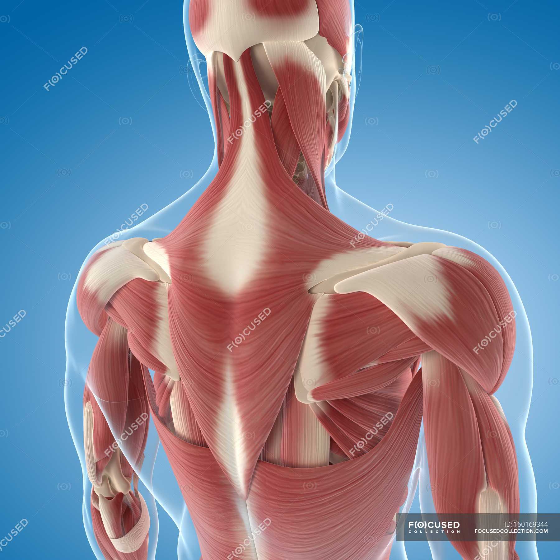
Upper Back Musculature Muscle Groups Anatomy Stock Photo 160169344 from st.focusedcollection.com Understanding lower back anatomy is key to understanding the root of lower back and hip pain. The bursa is a small sac of fluid that cushions and. These sections are cervical (neck), thoracic (upper and middle back), lumbar (lower back), and sacrum (tailbone). The complexity of this region means that dysfunction can occur either due to injury or progressive pain and degeneration. The lumbar and sacrum region make up the bone of the lower back anatomy. Back muscles anatomy here include the trapezius, latissimus dorsi, rhomboid and levator scapulae. The cause may be poor posture (such as forward head posture) or any type of irritation of the large back and shoulder muscles, including muscle strain or spasms. It is very stiff, and the thoracic spine has a limited range of motion.
See back muscle anatomy stock video clips.
Anatomy of the upper back. They originate from the vertebrae and insert into the scapulae. The rotator cuff is a collection of muscles and tendons that surround the shoulder, giving it support and allowing a wide range of motion. The back functions are many, such as to house and protect the spinal cord, hold the body and head upright, and adjust the movements of the upper and lower limbs. The upper back originates at the base of your neck, incorporates both shoulders and extends down to mid spine, including your ribs. This is my video about the muscles of the back. The bones of the chest and upper back combine to form the strong, protective rib cage around the vital thoracic organs such as the heart and lungs. This muscle is located on the upper portion of the back anatomy, underneath the trapezius. The back is the body region between the neck and the gluteal regions. The cause may be poor posture (such as forward head posture) or any type of irritation of the large back and shoulder muscles, including muscle strain or spasms. It is very stiff, and the thoracic spine has a limited range of motion. The superficial back muscles are situated underneath the skin and superficial fascia. Back muscles anatomy here include the trapezius, latissimus dorsi, rhomboid and levator scapulae.
The cause may be poor posture (such as forward head posture) or any type of irritation of the large back and shoulder muscles, including muscle strain or spasms. The main superficial muscles of the back are the following: The complexity of this region means that dysfunction can occur either due to injury or progressive pain and degeneration. It extends from the external protuberance of the occipital bone to the lower thoracic vertebrae and laterally to the spine of the scapula. This muscle is located on the upper portion of the back anatomy, underneath the trapezius.
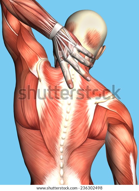
Anatomy Male Upper Back Pain Featuring Stock Illustration 236302498 from image.shutterstock.com Powerful muscles that move the head and arms attach to these bones as well. Understanding lower back anatomy is key to understanding the root of lower back and hip pain. They originate from the vertebrae and insert into the scapulae. The main superficial muscles of the back are the following: Upper back pain is most commonly caused by muscle irritation or tension, also called myofascial pain. The thoracic spine —also referred to as the upper back or middle back—is designed for stability to anchor the rib cage and protect vital internal organs within the chest. Both the deltoid and the trapezius are firmly attached to the spine of the scapula. The cause may be poor posture (such as forward head posture) or any type of irritation of the large back and shoulder muscles, including muscle strain or spasms.
Human musculature bodybuilding infographic muscular system vector human anatomy back muscle anatomy bicep male muscular anatomy human body anatomy female female anatomy muscle hamstrings muscle.
Both the deltoid and the trapezius are firmly attached to the spine of the scapula. Learn to draw the upper back muscles by understanding the anatomical details and forms. The bones of the chest and upper back combine to form the strong, protective rib cage around the vital thoracic organs such as the heart and lungs. Anatomy of the upper back. The trapezius muscle is a large superficial back muscle that resembles a trapezoid. This muscle is located on the upper portion of the back anatomy, underneath the trapezius. Vertebrae there are 12 vertebrae in the thoracic spine. The trapezius and latissimus dorsi muscles connect the upper limb to the vertebral column. The rhomboid muscle is activated as you bring and squeeze your scapula or shoulder blades back and together. The complexity of this region means that dysfunction can occur either due to injury or progressive pain and degeneration. The trapezius and latissimus dorsi muscles connect the upper limb to the vertebral column. The thoracic spine —also referred to as the upper back or middle back—is designed for stability to anchor the rib cage and protect vital internal organs within the chest. The back is the body region between the neck and the gluteal regions.
The lumbar and sacrum region make up the bone of the lower back anatomy. Try the injurymap exercise app now. The cervical spine supports the weight and movement of your head and protects the nerves exiting your brain. The iliocostalis muscles are furthest from the spine. The complexity of this region means that dysfunction can occur either due to injury or progressive pain and degeneration.
Back Pain Thoracic Joint Restriction from creeksidechiro.com The cause may be poor posture (such as forward head posture) or any type of irritation of the large back and shoulder muscles, including muscle strain or spasms. There is a set of muscles in the upper back (called the thoracic area) called the spinalis thoracis. It is like that for several reasons, all of which you can understand by looking at the anatomy of the thoracic spine. The iliocostalis muscles are furthest from the spine. Upper back pain is most commonly caused by muscle irritation or tension, also called myofascial pain. In the upper back region, the trapezius, rhomboid major, and levator scapulae muscles anchor the scapula and clavicle to the spines of several vertebrae and the occipital bone of the skull. They originate from the vertebrae and insert into the scapulae. The trapezius has upper, middle, and lower groups of fibers.
It comprises the vertebral column (spine) and two compartments of back muscles;
Back muscles anatomy here include the trapezius, latissimus dorsi, rhomboid and levator scapulae. The traps) the latissimus dorsi (a.k.a. The human spine is composed of 4 sections of vertebrae. There is a set of muscles in the upper back (called the thoracic area) called the spinalis thoracis. The bones of the chest and upper back combine to form the strong, protective rib cage around the vital thoracic organs such as the heart and lungs. The trapezius has upper, middle, and lower groups of fibers. Related posts of upper back muscle diagram anatomy muscle attachments. The iliocostalis muscles are furthest from the spine. The cervical spine supports the weight and movement of your head and protects the nerves exiting your brain. Anatomy of the upper back. This muscle is located on the upper portion of the back anatomy, underneath the trapezius. The trapezius and latissimus dorsi muscles connect the upper limb to the vertebral column. The complexity of this region means that dysfunction can occur either due to injury or progressive pain and degeneration.
Share :
Post a Comment
for "Upper Back Anatomy : Upper Back Chiropractor In Lexington Ky"

Post a Comment for "Upper Back Anatomy : Upper Back Chiropractor In Lexington Ky"